Spine surgery is traditionally an open surgery. This means the area being operated on is opened with a long incision to allow the surgeon to view and access the surgical site.
In recent years, technological advances have allowed more back and neck conditions to be treated with a minimally invasive surgical technique.
Because minimally invasive spine surgery (MISS), does not involve a long incision, it avoids significant damage to the muscles surrounding the spine and helps your doctor to see only the location where the problem exists in the spine.
The technique uses smaller instruments, which means the size of the surgical field (called exposure) is reduced, resulting in less pain after surgery and faster recovery.
Other benefits of minimally invasive spine surgery may include:
- Decreased blood loss
- Lower risk of muscle and soft-tissue damage
- Lower risks of infection
- Reduced postoperative pain
- Less pain medication use
- Shorter stays in the hospital
It may help alleviate some of your concerns to know that cervical disc replacement surgery is one such minimally invasive spine surgery. This procedure in an outpatient surgery.
In The Operating Room
The actual minimally invasive cervical disc replacement procedure may last a few hours. This is what may happen once the procedure begins:
Surgical Approach
- You are positioned on your back (supine) on a specialized, cushioned operating table.
- The front of the neck area is cleansed with a special solution to kill the germs on the skin.
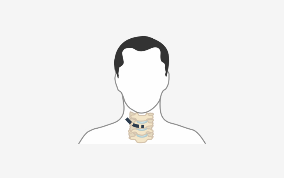
- The spine surgeon makes a small, horizontal skin incision (one-to-two-inch incision) on the side of the neck. Then, a tube is inserted through the incision and gently guide it through and between the muscles to your spine. A lighted microscope is used to help your doctor see the affected area and the cervical discs are visualized.
Disc Removal (Decompression)
- The spine surgeon makes an incision in the outer coating of the disc, and the soft inner core of the disc is removed using an operating microscope.
- The damaged disc is then extracted. This helps take the pressure off surrounding nerve roots and spinal cord and makes room for the new artificial disc.
Placement of the Artificial Disc Implant
After the discectomy (removal of the disc) has been completed, to prevent the vertebrae from collapsing and rubbing together, the artificial disc is inserted.
Depending on the disc design, the implant may be held in place with small screws.
Closure
- A drain is placed, and the incision is closed using absorbable sutures (stitches) under the skin.
- A small dressing is applied over the incision and a soft neck brace is applied. You are then taken to the Recovery Room.
Disclaimer – All information is for educational pursuit and information purposes only. It is not intended nor implied to be a substitute for professional medical advice. The viewer should always consult his or her healthcare provider to determine the appropriateness of the information for their own situation or if they have any questions regarding their medical condition, diagnosis, procedures, treatment plan, or other health related topics.